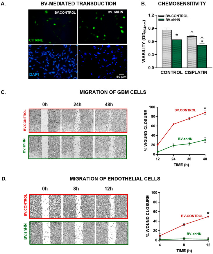
Figure 5.
BV-mediated silencing of HN in GBM cells. Murine GBM cells (GL26) were transduced with BV.Control or BV.shHN (750 pfu/cell) for 48 h. (A) Expression of the reporter gene (green) was assessed using fluorescent microscopy. (B) Transduced cells were incubated with cisplatin (5 μM) for 72 h and viability was assessed by MTT assay. * p < 0.05 vs. respective BV.Control, ^ p < 0.05 vs. respective control without cisplatin. ANOVA followed by Tukey’s test. (C) Murine GBM cells (GL26) were seeded until reaching confluence, transduced with BV.shHN or BV.Control (750 pfu/cells) and migration was evaluated at different time points using the wound assay. * p < 0.05 vs. BV.Control (nonlinear regression analysis). (D) EA.hy926 endothelial cells were seeded to confluence and incubated with conditioned medium from GBM cells transduced with BV-shHN. A wound assay was performed and the cell-free area was measured at different time points. * p < 0.05 vs. BV.Control (nonlinear regression analysis).

