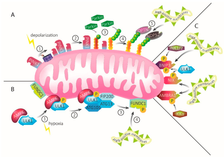
Figure 1.
Mechanisms of mitophagy. (A) PINK1–Parkin-mediated mitophagy. Under homeostatic conditions, PINK1 is constantly imported into mitochondria. Upon mitochondrial damage, import of PINK1 via the TOM complex and subsequent degradation is inhibited and the protein accumulates on the OMM (1). Accumulated PINK1 recruits Parkin (2), which ubiquitinates OMM proteins (3). Ubiquitination recruits the autophagy receptors OPTN and NDP52 (4). The autophagy receptors, in turn, recruit Atg8-positive membranes, leading to engulfment of the damaged mitochondria into mitophagosomal membranes (5). (B) ULK1–FUNDC1-mediated mitophagy upon hypoxia. Upon mitochondrial damage through hypoxia, AMPK activates ULK1 via phosphorylation (1), leading to the recruitment of FIP200, ATG13, and Atg101 (2). Together, they form the ULK1–AMPK complex. This complex interacts with the mitophagy receptor FUNDC1 that is abundant on mitochondrial membranes, leading to its phosphorylation (3). FUNDC1 possesses an LIR motif that recruits Atg8-positive membranes (4). These will eventually engulf the damaged mitochondria. (C) The mitophagy receptors BNIP3, NIX, and AMBRA1 possess LIR motifs which directly bind and recruit Atg8-positive membranes upon activation by signaling pathways, including ULK1, IKKα, and JNK1/2.

