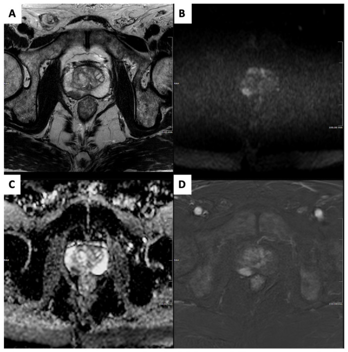
Figure 1.
Clinically significant prostate cancer in a 75-year-old biopsy-naïve subject who underwent magnetic resonance imaging (MRI) for raised prostatic-specific antigen level (7 ng/mL) and suspicious digital rectal examination. Prostate MRI showed a 13 mm PI-RADSv2.1 category 4 focus in the right mid-gland peripheral zone, demonstrating homogeneous moderate hypointensity on axial T2-weighted image (A); focal-restricted diffusion with marked hyperintensity on the high b-value image (B) and marked hypointensity on the apparent diffusion coefficient map (C); and focal, early enhancement on a digitally subtracted, fat-saturated T1-weighted image from the dynamic contrast-enhanced sequence (D). Targeted cores from transperineal biopsy showed a ISUP grade 2 prostate cancer.

