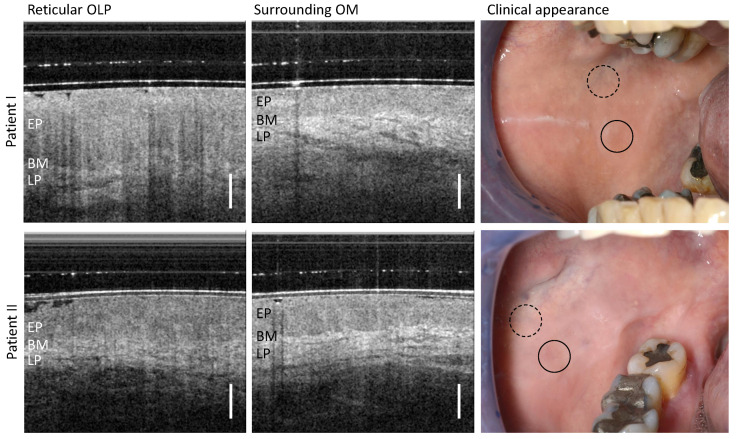
Figure 3.
OCT cross-sections of the center of reticular OLP (left column) and surrounding oral mucosa (OM) (middle column) of patient I (upper row) and II (lower row). Corresponding clinical appearance (right column) with marked zones of OCT scanning: OLP center (circle with black solid line) and surrounding OM (circle with black dashed line). Labels: EP: epithelium, BM: basement membrane, LP: lamina propria. Scale bar 500 µm.

