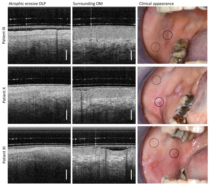
Figure 6.
OCT imaging of the center of the atrophic erosive form of OLP (directly adjacent to the open wounds) (left column) as well as more distant inconspicuous appearing regions of the buccal mucosa (middle column). Three patients (IX, X, XI) were examined, whose clinical findings with marked locations of OCT imaging are shown (right column): OLP center (circle with black solid line) and surrounding OM (circle with black dashed line). Scale bar 500 µm.

