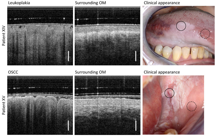
Figure 8.
(Upper row): Results of patient XIV clinically diagnosed with homogeneous leukoplakia of the ventral tongue. (Lower row): Results of patient XV visually diagnosed with homogeneous leukoplakia of the posterior buccal mucosa. The histologic findings of the biopsy taken immediately after OCT imaging revealed that this form of leukoplakia had degenerated into an oral squamous cell carcinoma (OSCC). Scale bar 500 µm. Upper row, leukoplakia (circle with black solid line) and surrounding OM (circle with black dashed line) lower row, OSCC (circle with black solid line) and surrounding OM (circle with black dashed line).

