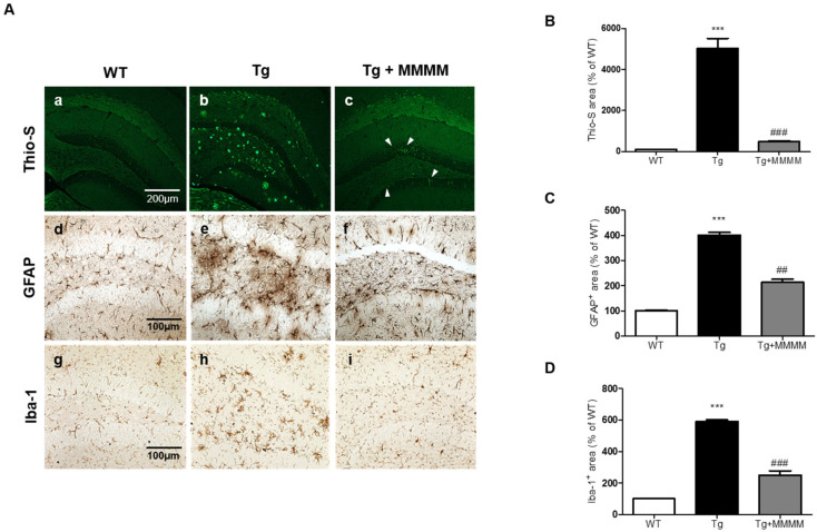
Figure 5.
Effects of MMMM on Aβ deposits and neuroinflammation in in vivo AD model. (A) Representative images of Thio-S fluorescence (a–c) and the IHC showing GFAP+ astrocytes (d–f) and Iba-1+ microglia (g–i) in different mice groups. (B) Thio-S-stained and (C) GFAP- and (D) Iba-1-immunostained areas were expressed using quantitative bar graphs. In all graphs, *** p < 0.001 vs. WT; ## p < 0.01 and ### p < 0.001 vs. Tg. Data are represented as the mean ± standard deviation. Thio-S, Thioflavin S; GFAP, glial fibrillary acidic protein; Iba-1, ionized calcium-binding adapter molecule1.

