Figure 4.
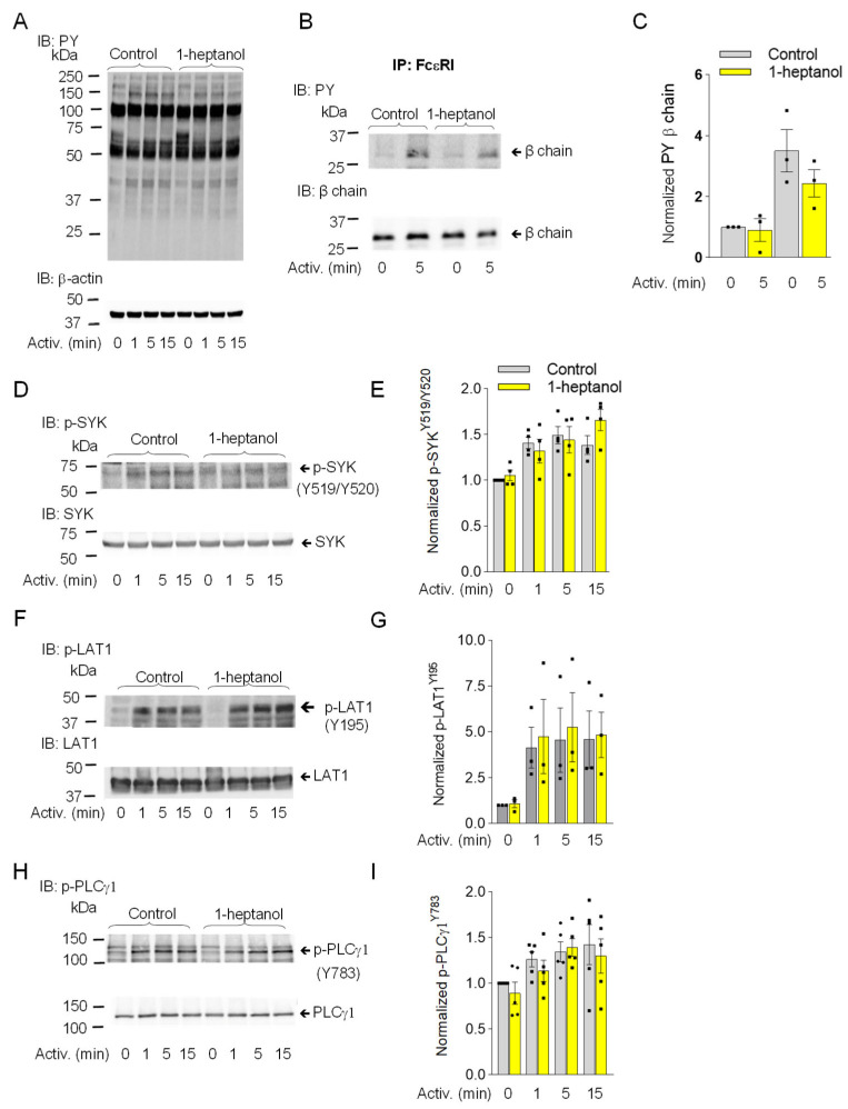
1-Heptanol does not affect tyrosine phosphorylation of global cellular proteins, FcεRI-βsubunit, or the SYK/LAT1/PLCγ1 axis in antigen-activated RBL-2H3 mast cells. (A) IgE-sensitized cells were treated for 15 min with vehicle (Control) or 2.5 mM 1-heptanol and then activated with antigen (TNP/BSA; 0.5 µg/mL) for the indicated time intervals in the absence or presence of 2.5 mM 1-heptanol. Whole-cell lysates were size-fractionated by SDS-PAGE and analyzed by immunoblotting (IB) with PY-20-HRP conjugate. For loading control, the blots were developed with an antibody specific for β-actin (n = 3). (B,C) The cells were treated for 15 min with vehicle (Control) or 2.5 mM 1-heptanol and then activated with antigen as in A for 5 min. The FcεRI complexes were immunoprecipitated (IP) and analyzed by IB with the PY-20-HRP conjugate. For loading controls, the blots were developed with FcεRI-β chain-specific antibody. The position of the FcεRI-β chain is indicated (n = 3). (C) Quantitative analysis of the phosphorylated FcεRI-β chains normalized to their amounts. Values indicate means ± SEM (n = 3). (D–I) RBL-2H3 cells were activated for different time intervals as in A, size-fractionated by SDS-PAGE, and analyzed by immunoblotting with phosphotyrosine-specific antibodies recognizing SYKY519/Y520 ((D,E); n = 4), LAT1Y195 ((F,G); n = 3), and PLCγ1Y783 ((H,I); n = 5). The antibodies specific to the corresponding proteins were used as loading controls. Representative immunoblots with the corresponding protein loading controls are shown in (D,F,H). The results from the quantification of the tyrosine-phosphorylated proteins in activated cells are normalized to signals in non-activated cells and loading control proteins (E,G,I). Values indicate means ± SEM calculated from the numbers above (n), denoting the numbers of biological replicates. Numbers on the left in (A,B,D,F,H) indicate the positions of molecular weight markers in kDa.

