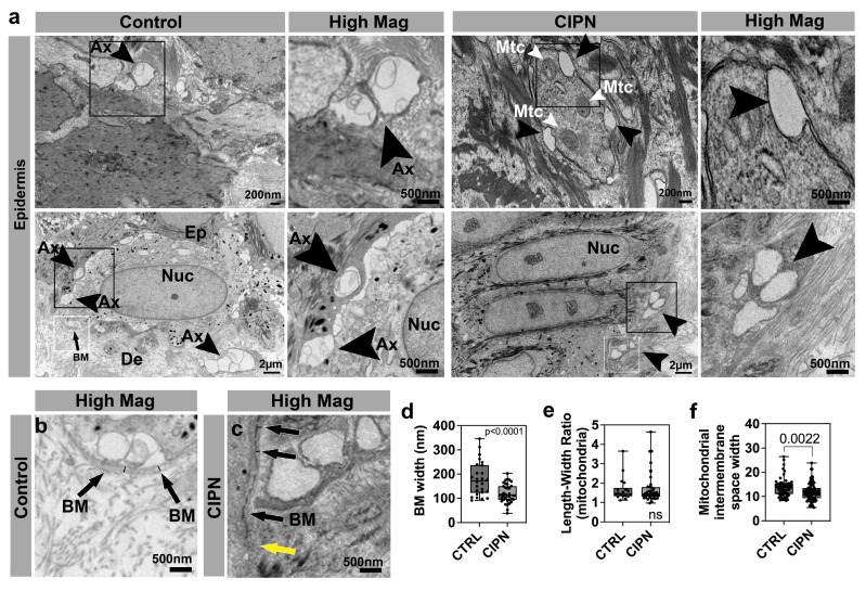
Figure 3.
Epidermal changes in CIPN patients. (a) Intraepidermal nerve endings (arrowheads) are abundant in control subject skin whereas gaps are present in the CIPN patient skin. (b) High magnification of control skin shown in (a) depicting the basement membrane (arrows) and width demarcated by the black lines. (c) High magnification of CIPN patient skin shown in (a) depicting the basement membrane (black arrows) and width demarcated by the black lines. The yellow arrow points to the less dense region of the basement membrane. (d) Quantification of the basement membrane width (white boxes in a) shows a reduction in the CIPN patient skin. (e) Length–width ratio (LWR) of mitochondria in epidermal keratinocytes shows no differences between the control subjects and CIPN patients. (f) Mitochondrial intermembrane space reduction in CIPN patients. Abbreviations: Ax: Axon, BM: Basement membrane, De: Dermis, Ep: Epidermis, Mtc: Mitochondria, Nuc: Nucleus.

