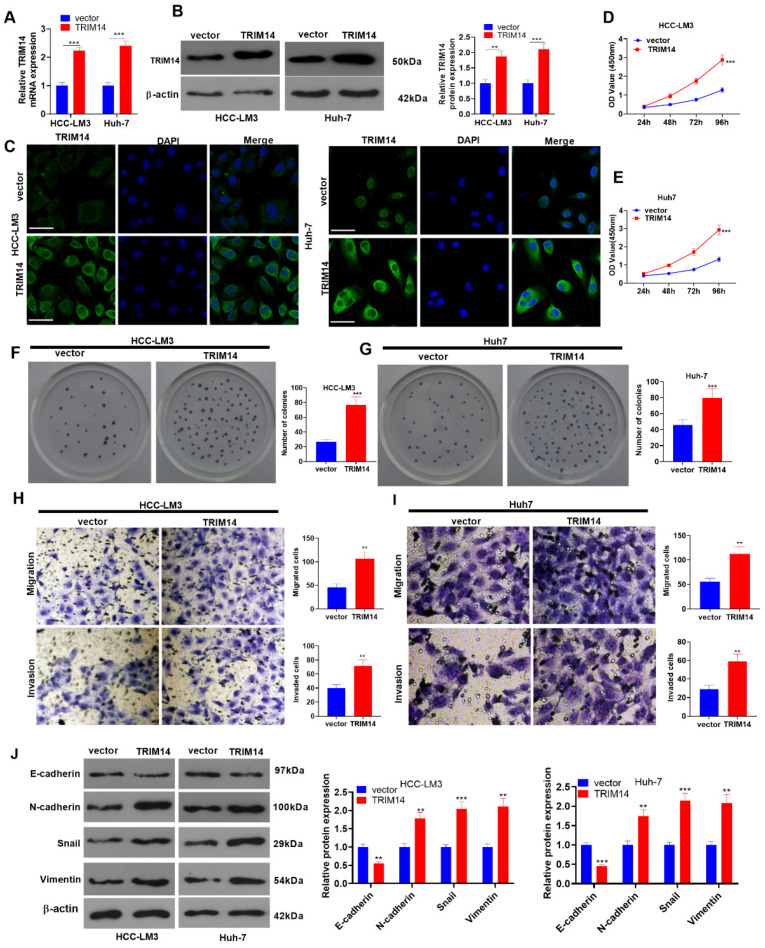
Figure 2.
To determine the role of TRIM14 overexpression in HCC cell proliferation and metastasis, HCC cells (HCC-LM3 and Huh-7) were transfected along with negative vector or TRIM14 overexpression plasmids. (A,B) TRIM14 expression in HCC cells was determined with qRT-PCR and Western blot. (C) Immunofluorescence was used to detect TRIM14 (green color) localization in HCC cells. Scale bar = 20 μm. The blue color shows DAPI. (D,E) CCK8 was used to examine HCC cell viability. (F,G) A clone formation assay was used to detect cell proliferation. (H,I) Transwell assays were used to monitor HCC cell migration and invasion. Magnification: 100×. (J) Western blot revealed the profiles of EMT-correlated proteins (including E-cadherin, N-cadherin, Snail, and Vimentin). ** p < 0.01 and *** p < 0.001 (vs. the vector group). N = 3.

