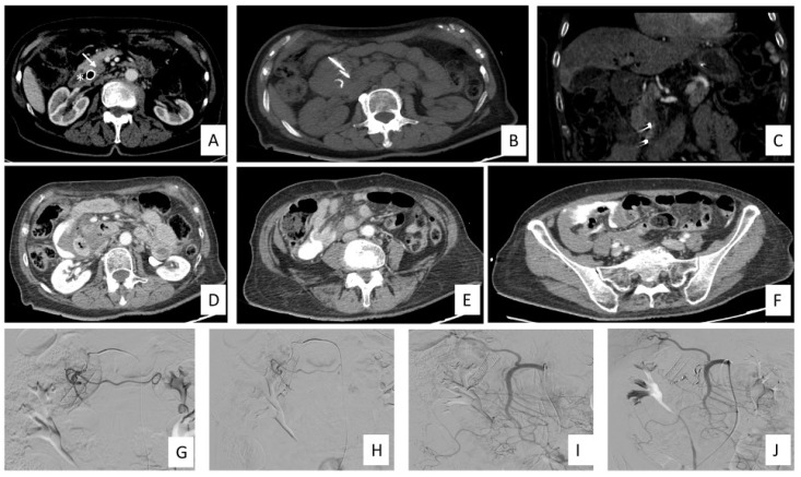
Figure 3.
IRE for LAPC treatment timeline and complication management in a 76-year-old female patient. (A) CECT in arterial phase demonstrates the presence of the LAPC in the head of the pancreas (white arrows) and biliary stent (asterisk) prior to IRE treatment. (B) Axial view of noncontrast scan shows two needle electrodes in situ. (C) Coronal view in arterial phase shows the two electrodes in situ. (D–F) CECT scan immediately post procedure shows the presence of intraabdominal hematic fluid and extravasation of contrast media without any visible source of bleeding. (G–J) Angiography of celiac trunk, SMA, GDA, right renal artery and phlebography of the inferior vena cava and right renal vein did not demonstrate any source of bleeding. A preventive endovascular embolization of GDA and PDA were made using 3, 4 and 5 mm micro coils. Abbreviations: contrast-enhanced CT (CECT), irreversible electroporation (IRE), gastroduodenal artery (GDA), locally advanced pancreatic cancer (LAPC), pancreatic duodenal artery (PDA), superior mesenteric artery (SMA).

