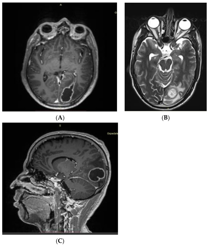
Figure 2.
(A) First axial contrast-enhanced T1-weighted NMR showing an oval-shaped enhanced lesion in the left occipital lobe with ipsilateral ventricular communication. (B) T2-weighted NMR showing ventricular communication of the lesion with T2 hyperintense fluid collection. (C) T1-weighted sagittal view of the abscess.

