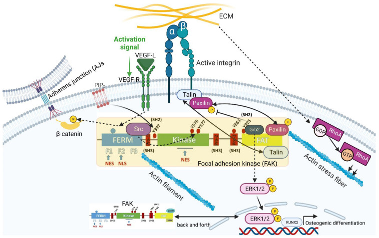
Figure 2.
Model of FAK activation mediated by different stimuli. FAs concomitant with actin stress fiber formation have been found to require RhoA activation in response to extracellular mechanical cues. FAK is activated upon integrin engagement. But first, clustering of FAK at the cell membrane lipid bilayer is induced by phosphatidylinositol-4,5-bisphosphate (PIP2) with the prior binding of PIP2 to the FAK basic region of FERM, leading to a partially opened conformation of the FAK molecule and to the exposure of the autophosphorylation site at tyrosine residue 397 (FAKp397). This partially opened conformation of FAK with FAK Y397 leads to the recruitment of Src molecules. These src molecules, in turn, lead to src-dependent phosphorylation of FAK Y576/577 within the kinase domain and prove that PIP2 is key to linking integrin signaling to FAK activation. FAK can also be phosphorylated at tyrosine residue 925 (FAK Y925). This phosphorylation is also carried out by growth-factor-receptor-bound protein 2 (Grb2). Grb2 can lead to the growth-factor-independent activation of the MAP kinase ERK2. Furthermore, the link between FAK and ERK1/2 can trigger osteogenic differentiation via the expression of TF Runt-related transcription factor 2 (RUNX2). Since the dissolution of FAs is putatively related to the phosphorylation of the FA component paxillin, it was found that this, in turn, is regulated by the impaired phosphorylation of FAK Y925. FAK is not only limited to the vertical contact area with the ECM but is also involved in mechanotransduction emerging from horizontally aligned cell–cell contacts such as adherens junctions (AJs). Here, FAK is able to specifically phosphorylate AJ-inherent β-catenin in response to vascular endothelial growth factor (VEGF) treatment. Since FAK can switch back and forth between the nucleus and cytoplasm, FAK has two nuclear export signal (NES) domains and one nuclear localization signal (NLS) domain. The schematic was created with BioRender.com.

