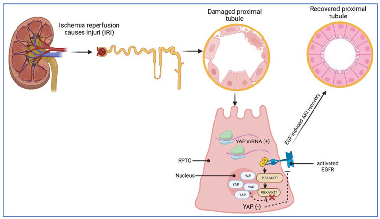
Figure 6.
Schematic illustration of YAP/TAZ in ischemia–reperfusion injury (IRI). Renal proximal tubular epithelial cells (RPTCs) isolated from renal mouse tissue displayed elevated levels of YAP mRNA in response to IRI. The same could be detected at the protein level in an IRI model system, in which YAP appeared predominantly with nuclear subcellular localization. Recovery from IRI-induced cell damage obviously requires the activation of the epidermal growth factor receptor (EGFR), which functions as an inductor of phosphatidylinositol 3 kinase (PI3K) protein signaling. Inhibition of EGFR and AKT1/PI3K revealed decreased levels of nuclear YAP and suggested EGFR-AKT1 signaling as a trigger of YAP activation. The schematic was created with BioRender.com.

