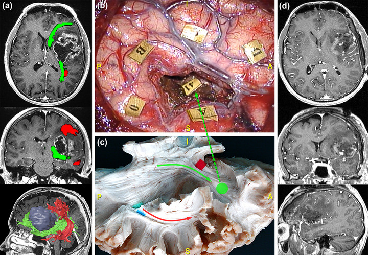
FIGURE 5.

Surgical case concerning a 42‐year‐old man who underwent resection of a high‐grade glioma located within the left dominant fronto‐insular region. (a) Preoperative magnetic resonance imaging (MRI) (from top to bottom: axial, coronal, and 3D‐sagittal sequences) combined with tractographic reconstruction of the left arcuate fasciculus (AF) (red) and inferior fronto‐occipital fasciculus (IFOF) (green). (b) Intraoperative picture showing the tumor's resection performed with “asleep‐awake‐asleep” technique. Tag 0 refers to speech arrest site at the cortical level, while stimulation in correspondence of tag 41, representing IFOF's frontal projections elicited anomia, semantic paraphasia, and perseveration during denomination task. (c) Dissection of the perisylvian region with Klingler technique. The specimen has been oriented according to the surgical perspective. The green circle corresponds to the site of direct electrical stimulation (DES) stimulation along the course of the frontal projections of the IFOF (green arrow). (d) Postoperative MRI showing the resection on the axial, coronal, and sagittal plane from top to bottom.
