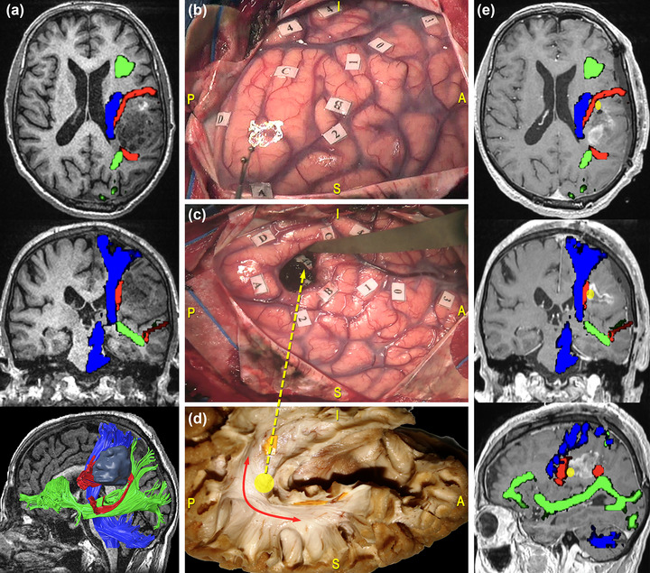
FIGURE 6.

Surgical case concerning a 69‐year‐old man who underwent resection of a high‐grade glioma located within the left dominant frontoparietal region. (a) Preoperative magnetic resonance imaging (MRI) (from top to bottom: axial, coronal, and 3D‐sagittal sequences) combined with tractographic reconstruction of the left arcuate fasciculus (AF) (red), the inferior fronto‐occipital fasciculus (IFOF) (green) and the corticospinal tract (CST) (blue). (b) Intraoperative picture showing the cortical mapping performed in awake condition with direct electrical stimulation (DES). Functional responses were found at the level of the precentral gyrus (PrCG), eliciting facial, and arm contractions (tag 1 and 2); of the ventral premotor cortex (VPMC), eliciting speech arrest (tag 0); of the pars triangularis (IFGtri), eliciting anomia (tag 3); of the posterior third of the superior temporal gyrus (STG), eliciting phonemic paraphasia (tag 4). (c) Intraoperative picture showing the results of subcortical mapping. Phonemic paraphasia was elicited at the level of the AF (tag 44). (d) Anatomical specimen of a left hemisphere oriented according to the surgical perspective and showing the AF course corresponding to the stimulation site (yellow circle and arrow) (e) Postoperative MRI (from top to bottom: axial, coronal, and sagittal sequences) combined with tractographic reconstruction of the left AF (red), the IFOF (green), and the CST (blue). The yellow circle corresponds to the site of elicitation of phonemic paraphasia (tag 44), matching with the AF course.
