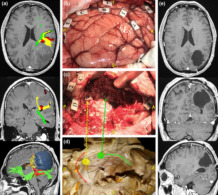
FIGURE 7.

Surgical case concerning a 41‐year‐old man who underwent resection of a high‐grade glioma located within the superior parietal lobule of the left dominant hemisphere. (a) Preoperative magnetic resonance imaging (MRI) (from top to bottom: axial, coronal, and 3D‐sagittal sequences) combined with tractographic reconstruction of the left arcuate fasciculus (AF) (red), the inferior fronto‐occipital fasciculus (IFOF) (green), and the temporoparietal component of the superior longitudinal fasciculus (SLF) (yellow). (b) Intraoperative picture showing the tumor's resection performed according to the “asleep‐awake‐asleep” protocol. Direct electrical stimulation (DES) allowed to identify eloquent cortical sites including: the postcentral gyrus (PoCG), eliciting right facial paresthesia when stimulated (tag 0); the supramarginal gyrus (SMG), eliciting speech arrest (tag 1) and anomia (tag 2 and 3) during naming tasks. (c) During tumor resection, subcortical mapping allowed to evoke semantic paraphasia at the level of the dorsal part of the IFOF (tag 41) and verbal apraxia during the denomination task, along the temporoparietal component of the SLF and the AF (tag 42). (d) Dissection of the perisylvian region with Klingler technique. The specimen has been oriented according to the surgical perspective. The colored tags correspond to the IFOF (green circle and arrow) and the temporoparietal SLF/AF (yellow circle, red, and yellow arrows). Postoperative MRI shows complete tumor resection.
