FIGURE 8.

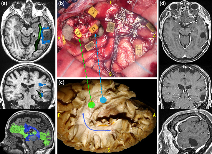
Surgical case concerning a 47‐year‐old woman who underwent resection of a high‐grade glioma located within the posterior third of the middle temporal gyrus (MTG) of the left dominant hemisphere. (a) Preoperative magnetic resonance imaging (MRI) (from top to bottom: axial, coronal, and 3D‐sagittal sequences) combined with tractographic reconstruction of the left arcuate fasciculus (AF) (blue) and inferior fronto‐occipital fasciculus (IFOF) (green). (b) Intraoperative picture showing the tumor's resection performed with “asleep‐awake‐asleep” technique. Direct electrical stimulation (DES) in awake condition allowed to identify eloquent cortical sites, including the ventral premotor cortex (VPMC), eliciting speech arrest (tag 0), and the ventral motor strip of the face (tag 1). During tumor's resection, the subcortical mapping allowed to identify the resection boundaries. These were established by eliciting phonemic paraphasia during the denomination task at the level of the anteroinferior portion of the AF (tag 42) and sematic paraphasia at the level of the IFOF (tag 43). (c) Dissection of the perisylvian region with Klingler technique. The specimen has been oriented according to the surgical perspective. The blue line corresponds to the AF stem and the light‐blue and green circles and arrows to the site where the IFOF and the AF were stimulated, respectively. (d) Postoperative MRI showed the complete tumor resection on the axial, coronal, and sagittal planes from top to bottom.
