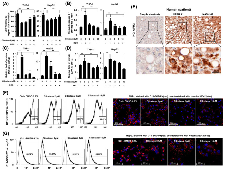
Figure 6.
Cilostazol attenuated RBC-induced ferroptosis in THP-1 and HepG2 cells. (A–D) Coculture experimental system. (A) Cell viability assay. (B) Quantification of the ratio of cleaved caspase-3 to procaspase-3. (C,D) Quantification of western blotting for the ratio of AIFM2 (C) and GPX4 (D) to the loading control. (E) Immunohistochemistry of AIFM2 in liver tissue from human NASH patients. (F,G) Flow cytometry assay and confocal microscopy of THP-1 (F) and HepG2 (G) cells stained with BODIPYTM 581/591 C11. Confocal microscopy stained with BODIPYTM 581/591 C11 (red) and counterstained with Hoechst 33342 (blue). ** p < 0.01 according to Scheffe’s test.

