Figure 1.
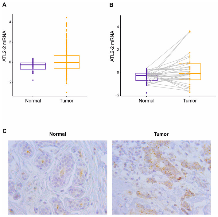
ATL2-2 is more highly expressed in breast tumors than normal breast tissue. (A) ATL2-2 transcript levels were compared between breast tumors and normal (non-neoplastic) breast tissue samples from Cohort 2 using a Student’s t-test (263 tumors vs. 36 normal tissue), p = 6.0 × 10⁻⁶, and (B) in 36 tumors and their corresponding adjacent normal tissue using a paired t-test, p = 0.05. Tumors are depicted in yellow and normal tissue in blue. (C) ATL2 protein expression was detected by ATL2 antibody in tumor cells (granular brown cytoplasmic stain) and adjacent normal cells (faint brown). A representative figure from immunohistochemical analysis of 13 tumor–normal pairs. The magnification was 40x. ATL2 protein expression was mostly observed in the cytoplasm in granules and as a diffuse stain.

