Figure 3.
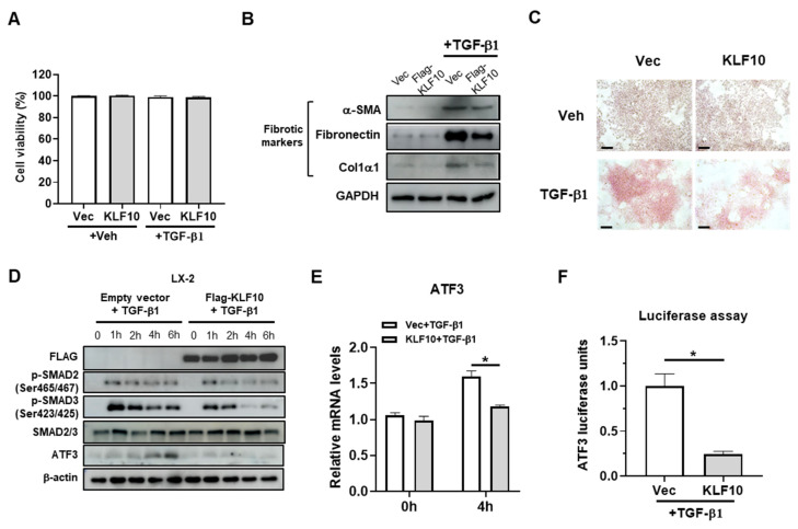
KLF10 inhibits TGF-β1-induced activation of LX-2 cells. LX-2 cells were transfected with pcDNA-KLF10 for 48 h and further treated with 10 ng/mL TGF-β1 for another 24 h. (A) Cell viability was determined by MTT assays. (B) Protein levels of α-SMA, fibronectin, and Col1α1 were determined by Western blotting. (C) Collagen accumulation was evaluated using Picro-Sirius red staining. Collagen, red. Scale bar = 100 μm. (D) LX-2 cells were transfected with pcDNA-KLF10 for 48 h and treated with TGF-β1 for the indicated times. The levels of p-SMAD2, p-SMAD3, SMAD2/3, KLF10, and ATF3 were determined using Western blotting. β-actin was used as an internal control. (E) LX-2 cells were transfected with pcDNA-KLF10 for 48 h and further treated with 10 ng/mL TGF-β1 for another 4 h. mRNA levels of ATF3 were determined by qPCR. Data are expressed as means ± SEM. (F) LX-2 cells were transfected with pcDNA-KLF10 for 48 h and further treated with 10 ng/mL TGF-β1 for 24 h. ATF3 promoter activity was determined by a luciferase assay. * p < 0.05.

