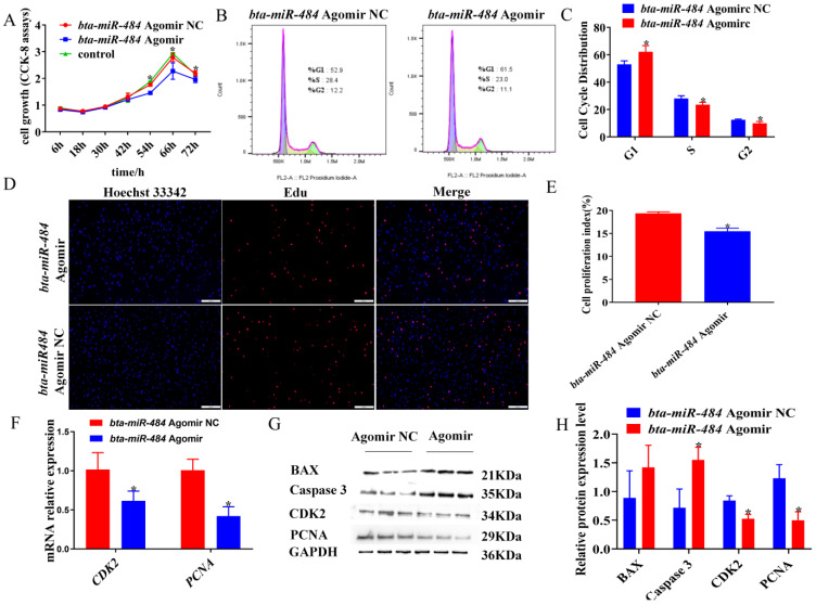
Figure 2.
Bta-miR-484 agomir inhibited the proliferation of preadipocytes. (A) CCK-8 assay to determine adipocyte viability. (B,C) Cell cycle phase analysis of adipocytes by flow cytometry. (D,E) Adipocyte proliferation was examined by EdU immunofluorescent staining. Red represents EdU staining; blue represents cell nuclei stained with Hoechst 33342. Scale bar: 500 μm. (F) Relative mRNA expression of CDK2 and PCNA genes in preadipocytes 48 h after transfection with a bta-miR-484 agomir or bta-miR-484 agomir NC. (G,H) Protein expression levels of CDK2, PCNA, Bcl-2-associated X protein (BAX), and cysteine-dependent aspartate-specific proteases 3 (Caspase 3) were detected in bta-miR-484 over-expressing adipocytes using WB. GAPDH was used as an internal reference. Data are presented as mean ± SD. n = 3. * p < 0.05.

