Figure 1.
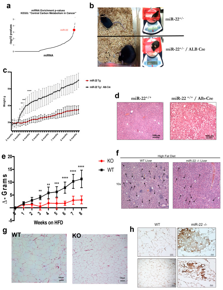
Role of miR-22 in metabolism and obesity. (a) The central carbon metabolism pathway is enriched in miR-22 targets. Dot plot depicting -log10 Fisher’s exact test p-values for all human miRNAs (n = 1077) in TarBase v8 database of experimentally supported miRNA interactions [29156006]. miR-22 is marked in red. (b) Representative comparison between miR-22+/− and miR-22+/−/Alb-Cre littermates at 8 months of age; mice were fed with regular chow for the entire length of the experiment. miR-22Tg/Alb-Cre mice weighed over two times more than their respective littermates without Cre. (c) Comparison between miR-22+/+ transgenic mice with or without Albumin Cre expression. Mice fed with regular chow showed a striking difference between the two genotypes; miR-22+/+-Alb-Cre mice gained much more weight than miR-22+/+ and started to become obese (>40 g) around 6 months of age. The entire colony was over the obesity threshold by 9 months of age (n = 6 per cohort). (d) Livers from miR-22+/+ transgenic mice with or without Albumin Cre were harvested at 10 months of age. H&E staining revealed marked liver steatosis in miR-22+/+-Alb-Cre mice fed regular chow, while there was no sign of liver steatosis in livers from WT mice. (e) HFD-fed miR-22 deficient mice failed to gain weight compared to WT littermates, suggesting a role of miR-22 in diet-induced obesity (n = 5 per cohort). (f) Livers from miR-22−/− mice were harvested after 8 weeks on an HFD. H&E staining revealed a strong protective effect against hepatic steatosis in the mice lacking miR-22. (g) After 8 weeks on an HFD, mice were sacrificed and tissues harvested. WAT from miR-22-KO and WT mice stained with H&E showed a significant difference in adipocyte size, which was much smaller in miR-22 KO mice compared to their WT littermates. (h) WAT from WT and miR-22−/− mice fed an HFD for 8 weeks was stained for UCP-1; miR-22-deficient mice showed strong staining, suggesting brownization of WAT in miR-22 null mice. *, p < 0.05; **, p < 0.01; ***, p < 0.001; ****, p < 0.0001.

