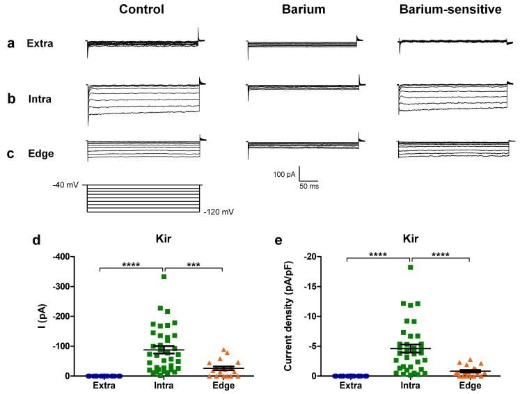
Figure 3.
Hyperpolarizing protocol for investigating the inward rectifier current. (a,b) Experimental traces obtained using a hyperpolarizing voltage step protocol (bottom) in the cells recorded extra-scratch (a), intra-scratch (b), and at the edge (c) of the wounded area in the control conditions (left) and after perfusion with barium 1 mM (center). The barium-sensitive current was obtained by digital subtraction (right). (d) Scatter plot of the amplitude of the KIR current measured at −120 mV from individual cells at different locations of the scratch area. (e) Scatter plot of the current density of the KIR current measured at −120 mV from individual cells at different locations of the scratch area. p-values: *** p < 0.001, and **** p < 0.0001.

