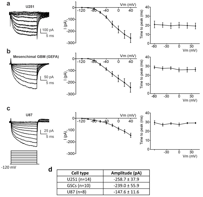
Figure 5.
Detection of the inward current at the edge of the wounded area in different cell lines. (a–c) Left: experimental traces elicited by the application of a depolarizing protocol of the voltage steps (shown at the bottom) from a holding potential of −120 mV to + 40 mV in U251 (a), mesenchymal GSCs (b), and U87 cells (c). Center: mean I–V relationship for U251 (n = 14 cells, (a)), mesenchymal GSCs (n = 10 cells, (b)), and U87 (n = 8 cells, (c)). Right: average inward current activation time to peak measured at different test potentials for U251 (n = 14 cells, (a)), mesenchymal GSCs (n = 10 cells, (b)), and U87 (n = 8 cells, (c)). (d) Averaged amplitudes of the inward current at +40 mV, measured by means of a depolarizing step from a holding potential of −120 mV, recorded in different cell lines at the edge of the wounded area.

