Figure 6.
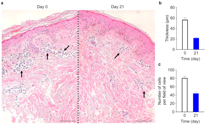
Corneum stratum and lymphocyte infiltration at the dermis–epidermis interface in skin lesions of one subject before and after topical application of SPM. (a) Histopathological examination of treated skin as assessed by hematoxylin-eosin (H & E) staining. Lymphocyte infiltration (pockets of nuclei, black arrows) at the dermis-epidermis interface. 200× objective. Scale bar is 100 μm. (b) Stratum corneum thickness before and after 3 weeks of SPM treatment. (c) Counts of lymphocytes infiltrating the epidermis–dermis interface before and after 3 weeks of SPM treatment.

