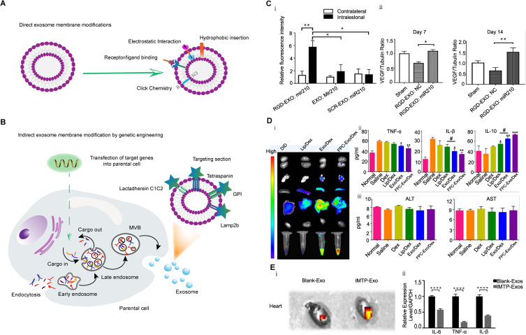
Figure 5.
Strategies and applications of exosome membrane modification. (A) Direct modification of exosome membrane strategies. (B) Indirect modification of exosome membrane strategies by genetic engineering. (C) Targeting peptide c (RGDyK)-modified exosomes loaded with miR-210 for the treatment of cerebral ischemia models.184 Reprinted from Zhang H, Wu J, Wu J, et al. Exosome-mediated targeted delivery of miR-210 for angiogenic therapy after cerebral ischemia in mice. J Nanobiotechnology. 2019;17(1):29. http://creativecommons.org/licenses/by/4.0/. (i) Fluorescence intensity of GRD-modified exosomes and unmodified exosomes loaded with miR-210 in a brain ischemia-reperfusion model in the lesioned (intralesional) and matched non-lesioned (contralateral) areas 6 hours after intravenous injection, *P < 0.05, **P < 0.01. (ii) VEGF expression in the lesion area at 7 and 14 days after reperfusion and administration of RGD-exo:NC or RGD-exo:miR-210, *P < 0.05, **P < 0.01. (D) FPC-modified exosome for RA-targeted therapy.25 Reprinted from Yan F, Zhong Z, Wang Y, et al. Exosome-based biomimetic nanoparticles targeted to inflamed joints for enhanced treatment of rheumatoid arthritis. J Nanobiotechnology. 2020;18(1):115. http://creativecommons.org/licenses/by/4.0/. (i) In vivo imaging of plasma and organs 24 hours after intravenous injection of Dex-loaded liposomes, Dex-loaded exosomes, and Dex-loaded FPC-modified exosomes. (ii) Serum levels of inflammatory cytokines (TNF-α, IL-1β and IL-10) in normal and treated CIA mice, *p < 0.05, **p < 0.01, ***p < 0.001, #p < 0.05. (iii) Serum levels of ALT and AST in normal and treated CIA mice. (E). IMTP-Exosomes for Myocardial Ischemia Targeted Therapy.185 Reprinted from Wang X, Chen Y, Zhao Z, et al. Engineered Exosomes With Ischemic Myocardium-Targeting Peptide for Targeted Therapy in Myocardial Infarction. J Am Heart Assoc. 2018;7(15):e008737. http://creativecommons.org/licenses/by/4.0/. (i) Fluorescence imaging of DiR-labeled blank-Exos and IMTP-Exos in a mouse model of myocardial ischemia after 72h of intravenous injection. (ii) Expression levels of inflammatory factors (TNF-α, IL-1β and IL-6) after IMTP-Exos and blank-Exos treatment, ****p < 0.001.

