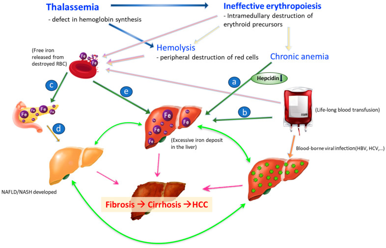
Figure 1.
Mechanisms of HCC development in thalassemia. a (in blue circle): Thalassemia is characterized by ineffective erythropoiesis and has low hepcidin levels and iron overload. b (in blue circle): Excessive iron from senescent RBC. c (in blue circle): Excessive iron deposit in endocrine organ results in pancreatic exocrine insufficiency which causing type 1 DM. d (in blue circle): high fat/fructose/glucose diet, and sedentary lifestyle. e (in blue circle): Excessive iron also deposits into liver. Blue arrow: Thalassemia causes premature destruction of RBC precursor and hemolysis owing to deformed RBC. Light pink arrow: Hemolysis, ineffective hematopoiesis, and chronic transfusions lead to premature death of RBC. Free iron is released accordingly. Bright pink arrow: NASH, iron overload and chronic hepatitis viral infection all accelerate the process of fibrosis to cirrhosis and worst but not always consequential, to HCC. Green arrow: NASH, iron overload and chronic hepatitis viral infection can aggravate each other and make the progress of liver fibrosis early-onset, more inevitable, and severe.

