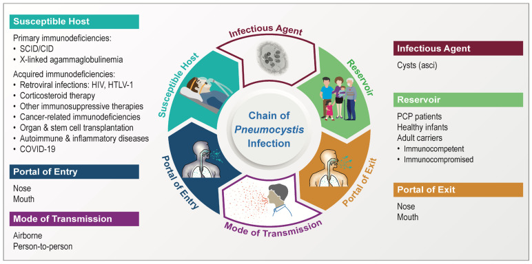
Figure 1.
Pneumocystis infection chain. The infectious agent is exemplified by the cyst (also known as ascus) of P. murina in infected mouse lungs revealed by transmission electron microscopy at a magnification of 5000×. The cyst is characterized by a thick wall with double electron-dense layers enclosing eight intracystic bodies or spores. For the primary immunodeficiencies (under the Susceptible Host), only a limited number of conditions are listed as examples to enhance visual clarity, with more details described in Section 4.5.

