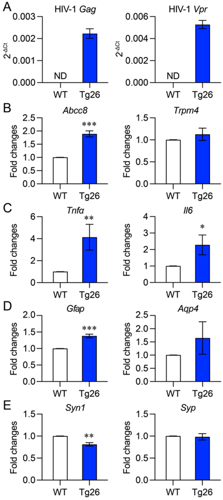
Figure 5.
Comparison of proinflammatory and synaptic gene transcription in the cortex and hippocampus of WT and Tg26 mice. (A) Verification of transgenic HIV-1 gene expression in the Tg26 and WT mice of mixed sexes was performed by qPCR. Representative transcriptional gene expression of proinflammatory markers Abcc8 (which encodes SUR1 protein) and Trpm4 (B), Tnfα and Il6 (C) astrocyte activation (Gfap and Aqp4) (D), and neuronal synaptic function markers Syn1 and Syp (E) were measured in the cortex and hippocampus brain tissue of WT and Tg26 mice. The gene expression profiles are presented as relative ratio to house-keeping Gapdh gene (as indicated by 2−ΔCT value) in each sample (A) or relative fold change to WT (B–E). Gapdh gene was used as an internal control. The data are presented as Mean ± SD. An unpaired t-test was used to determine statistical significance between the two testing groups, with * p < 0.05, ** p < 0.01, and *** p < 0.001 indicating the levels of significance.

