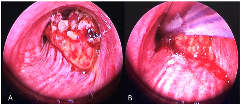
Figure 3.
The figure shows a mid-trachea obstructive tumor leaving a very small airway lumen. (A) The FOT tube was passed already and cuffed below the stenosis. The upper airway was excluded from the distal airway below the cuff. The rigid bronchoscope was placed, and the operative phase could be started (B).

