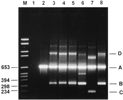
FIG. 3.
Agarose gel electrophoresis of PCR-amplified DNA fragments of the pbp2B gene from S. pneumoniae. Lane M, molecular size marker (in base pairs). Lane 1, negative control; lane 2, penicillin-susceptible S. pneumoniae. Primer combinations are as follows: R1 + P5 + P6 (lane 3), R3 + P5 + P6 (lane 4), R1 + R3 + P5 + P6 (lane 5), R2 + P5 + P6 (lane 6), R4 + P5 + P6 (lane 7), and R2 + R4 + P5 + P6 (band C is poorly visible) (lane 8). (A) A 682-bp species-specific product arising from amplification with primers P5 and P6. (B) A 328- to 334-bp products arising from amplification with primers R1 to R3 and P6. (C) A 214-bp product arising from amplification with primers R4 and P6. (D) Amplification products produced as a result of annealing between a resistance product(s) and the 682-bp product and which are subsequently extended by Taq DNA polymerase to produce a larger product (±900 to 1,000 bp).

