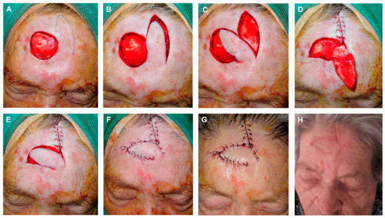
Figure 4.
(A) Final defect with free margins down to the periosteum. Design of the finger-shaped transposition flap. (B,C) Flap sculpted on a deep plane, lifted and displaced. (D) Donor area sutured with clamps. Reduction in the defect height by a subcutaneous guitar-string suture. These vertical dermal–subcutaneous sutures begin at the depth of the wound and roll toward the surface, reenter the opposite side of the wound in the dermis and roll deep, thus creating uniform tension across the wound and significantly decreasing the size of the defect. (E) Flap pulled into place. De-epithelialization of the turning point to prevent a bulge fold; the epidermis and superficial dermis are removed to avoid this fold, but the rest of the layers maintained to ensure vascular supply of the FMTF. (F) Immediate result after closure with 4/0 polyglactin and 6/0 silk sutures. (G) Appearance 24 h after surgery. (H) Appearance one month after surgery.

