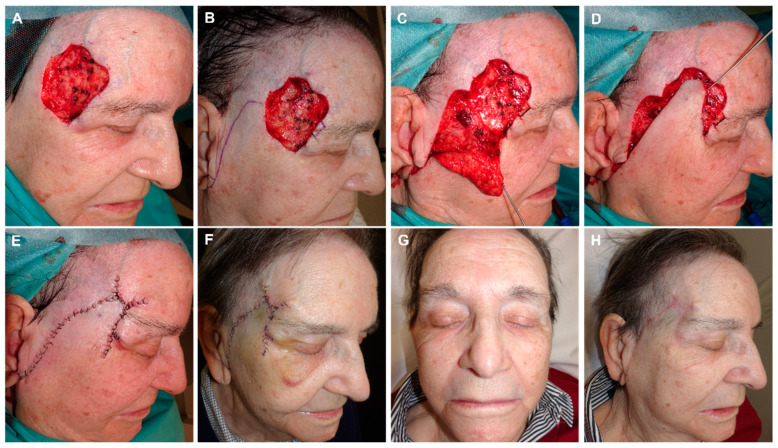
Figure 8.
(A) Basal cell carcinoma on the temple. Initial defect with a positive lateral margin after 3D histology study. (B) Margin demarcated for extension and closure design using an advancement rotation flap from preauricular skin with an infra-lobular Burow’s triangle. (C) Flap lifted on the subcutaneous plane, (D) and pulled into place with a dissecting hook. (E) Design of three Burow’s triangles to reduce the size of the defect. Immediate result. (F) Appearance one week later. (G,H) Final aspect (front and lateral) 4 months after surgery.

