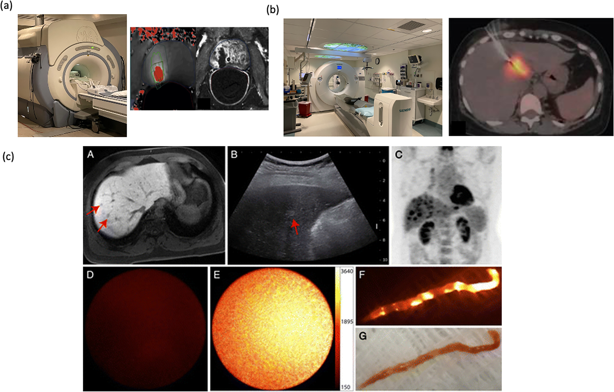
Figure 7.

Uncommon clinical imaging modalities. a) MRI machine, and MR guided focal ultrasound ablation showing MR thermometry and area of ablation. Adapted with permission from Napoli et. al. Copyright European Urology[105] b) PET- CT, PET guided biopsy of a liver lesion, Adapted from Govindarajan, et. al. Creative Commons License [108]. c) Use of near infrared imaging to confirm biopsy specimen subpart A shows lesion on MRI, subpart B shows on ultrasound, subpart C shows FDG avidity, sub-part D shows no fluorescence on normal liver, sub-part E shows fluorescence during active guidance in sub-part E, and fluorescence on external imaging, sub-part F, and corollary visuals in sub-part G. Images reproduced with permission from Sheth et. al. Radiology 2014 Copyright Radiology.[109]
