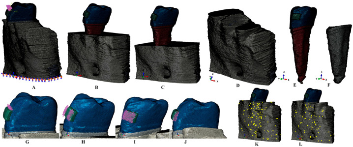
Figure 1.
(A) 2nd lower right premolar model with intact periodontium, and applied vectors (encastered model base and extrusion loads); (B) 4 mm bone loss; (C) 8 mm bone loss; (D) bone structure (with cortical and trabecular components); (E) tooth model with bracket, enamel, dentin and neuro-vascular bundle, (F) intact PDL; applied vectors: (G) intrusion, (H) rotation, (I) tipping, (J) translation; (K) element warnings of the cortical bone component; (L) elements warnings of the cortical component.

