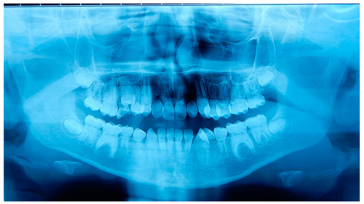
Figure 3.
Orthopantomography of patient A. This radiograph was made at the moment of the dental check-up of patient A (12 years old), showing the typical abnormalities of the DD-I affect the roots (short with apical constrictions) and the pulp (pulp chamber and canals obliteration, half-moon shaped pulp chamber remnants) and the rootless permanent teeth (the upper lateral incisors and the lower first premolars).

