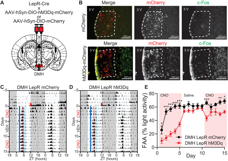
Fig. 6. Pre-FAA activation of DMHLepR neurons suppresses the development but not maintenance of food entrainment.
(A) Schematic diagram illustrating bilateral injection of AAV-hSyn-DIO-hM3Dq-mCherry or AAV-hSyn-DIO-mCherry to the DMH of LepR Cre mice. (B) Representative images showing the expression of mCherry (top) or hM3Dq-mCherry (bottom) in DMHLepR neurons and c-Fos response 2 hours after CNO injection. (C and D) Representative actograms of (C) DMHLepR mCherry and (D) DMHLepR hM3Dq mice on SF that received CNO (0.3 mg/kg; SF days 1 to 5), saline (SF days 6 to 10), and CNO (0.3 mg/kg; SF days 11 to 15) injection at ZT2.5. Shading color scheme is described in Fig. 3C. See fig. S9 (E and F) for actograms of all animals. (E) Quantification of FAA. Pink-shaded areas indicate days with CNO injection. No shading indicates saline injection. Repeated-measures two-way ANOVA with Bonferroni post hoc comparison; n = 8 to 9 per group; Fvirus (1,15) = 46.24, P < 0.0001. Data are represented as means ± SEM. *P < 0.05; **P < 0.01; ***P < 0.001.

