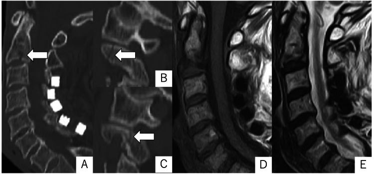
Figure 1. Sagittal CT images (A: midline, B: right, C: left) and magnetic resonance (MR) images (D: T1W, E: T2W) obtained in the early days after the injury.
CT images showed fracture lines in the C2 pedicle and vertebral body (arrows). MR images showed signal changes in the vertebral body, but no evidence of disc or ligament injury.
T1W: T1-weighted; T2W: T2-weighted.

