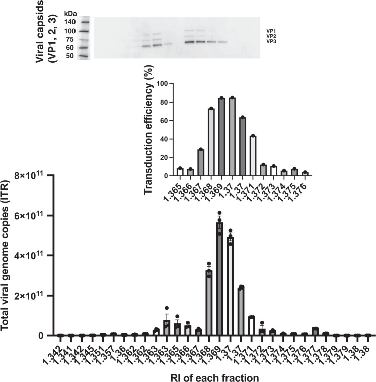
Fig. 2. AAV vectors separated for full-genome, intermediate, and empty particles were analyzed by genome copies, transduction efficiency, and AAV capsid proteins.
(Top) AAV capsids were detected by western blotting using anti-VP1 (82 kDa), VP2 (67 kDa), and VP3 (60 kDa) antibodies. (Middle) AAV transduction efficiency was evaluated using ZsGreen1 expression in transduced 293EB cells (n = 1, means ± SEM). (Bottom) AAV genome copies were measured by quantitative polymerase chain reaction (qPCR) using inverted terminal repeat (ITR)-targeting primers (n = 3, means ± SEM). Experiments were repeated six times in a single run.

