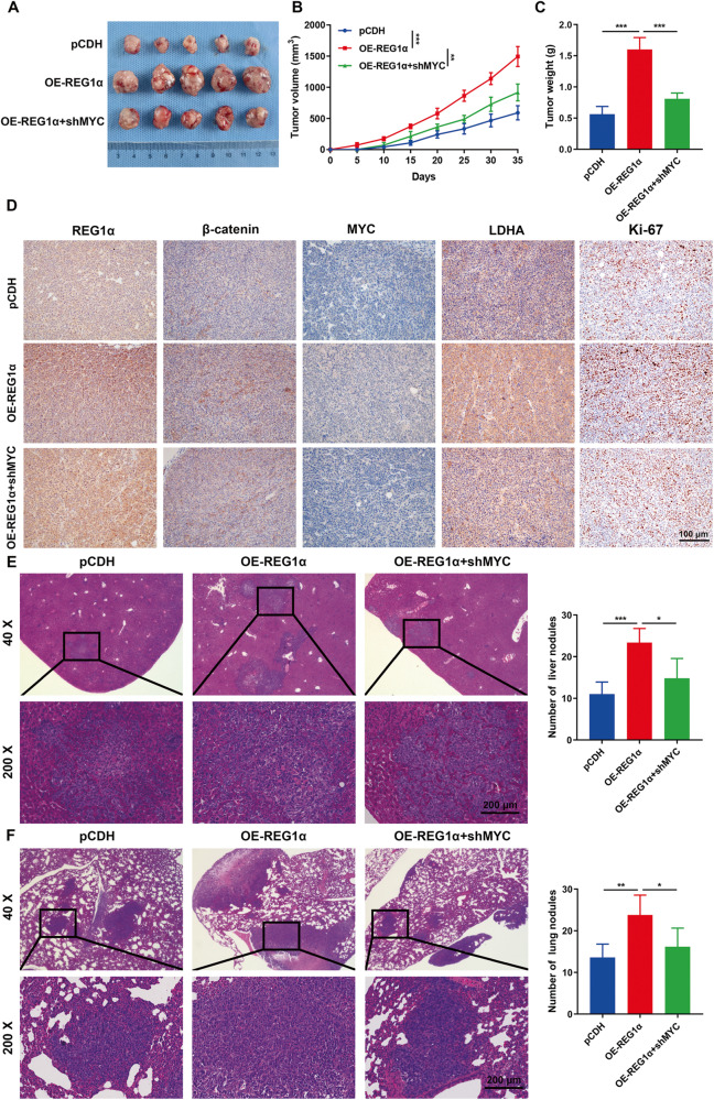
Fig. 7. REG1α facilitates tumorigenesis and metastasis of CRC cells in a MYC-dependent manner.
A Images of tumors formed in nude mice bearing control SW620 cells and REG1α-overexpression cells with or without MYC inhibition. B Tumor volumes were monitored every five days and tumor growth curves were shown. C The weight of xenograft tumors between different groups were measured at the endpoint of experiment. D Representative IHC images of REG1α, β-catenin, MYC, LDHA and Ki-67 staining in serial sections of tumor tissues isolated from subcutaneous models. E Representative images of H&E staining of liver sections in nude mice (n = 5 per group) after intrasplenic injection of SW620 cells. Statistical analysis of metastatic nodules was evaluated. F SW620 cells with indicated plasmids were injected into the tail veins of nude mice and the numbers of metastatic foci in the lung were quantified. Scale bar = 200 μm. Data were expressed as the mean ± SD. Significant differences were shown by *P < 0.05, **P < 0.01, and ***P < 0.001.

