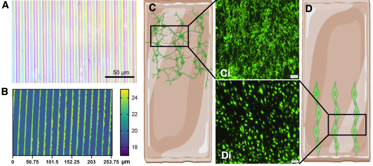
FIG. 4.
Depositing cells on the magnetic cantilevers. (A) Optical micrograph of the micropatterning stamp, with a pattern line of 10 μm wide with a spacing of 5 μm. (B) Topography of the stamped groove pattern on the scaffold. (C,D) Illustration of micropattern hydrogels with (C,Ci) aligned differentiated SH-SY5Y cells and (D,Di) endothelial cells. Scale of the confocal imaging, 100 μm.

