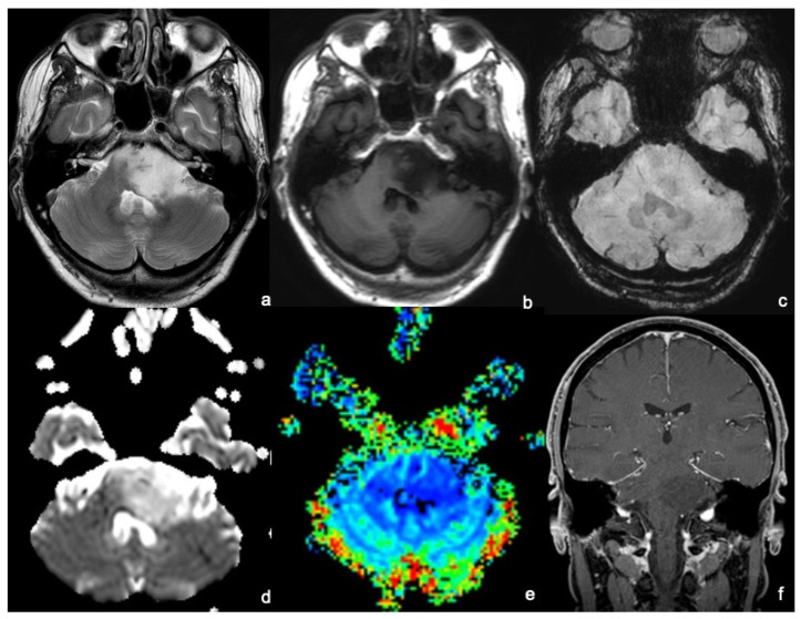
Figure 1.
An MRI of a 45-year-old man presenting with a long-lasting headache and dizziness related to a diffuse intrinsic low-grade glioma. MRI shows a bulky lesion of the left pons extending: anteriorly, partially obliterating the prepontine cistern and contacting the patent basilar; posteriorly to invade the left–middle cerebellar peduncle and to compress the IV ventricle; and medially to the right hemipons. The lesion appears to be inhomogeneously hyperintense on T2WI (a) and hypointense on T1WI (b), without hemorrhagic foci on SWI (c), and without diffusion restriction foci on the ADC map (d). After gadolinium administration, the tumor does not enhance on T1WI (f), and there is no increased perfusion on the rCBV map (e). The anatomopathological analysis demonstrated that the lesion was a grade 2—2021 WHO classification of brain tumors.

