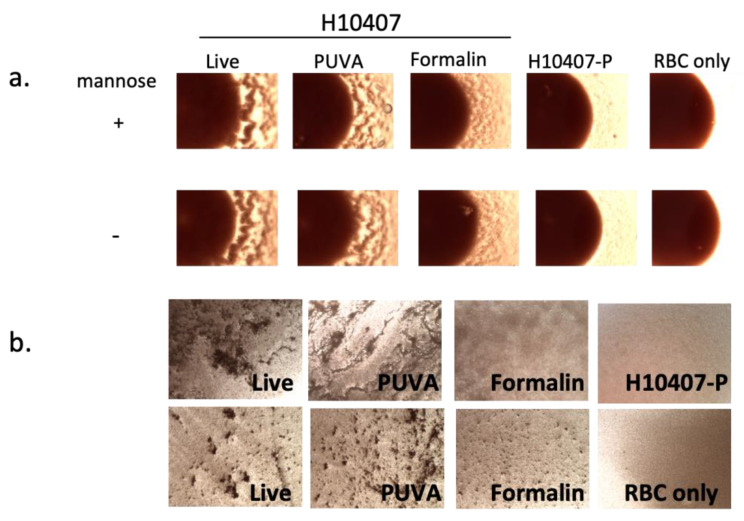
Figure 3.
MRHA activity of live, PUVA- and formalin-inactivated ETEC. Bacteria were mixed with (a). human Type A RBC ± mannose in a 96-well round bottom plate or (b). with human Type A RBC + mannose on glass slides as described in methods. Photomicrographs captured under visible light with an RGB filter at 4× magnification using an EVOS M5000 Imaging System depict (a). partial wells showing agglutination visible at RBC pellet edges after 24 h of settling or (b). agglutination visible as clumping on slides after 1 h; 2 fields are shown for each sample in (b). Controls were RBC only and H10407-P [43], which lacks the pCS1 virulence plasmid encoding CFA/I [46]. Well and slide assays were performed at least 3 times with similar results.

