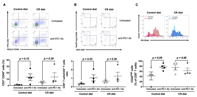
Figure 3.
Effect of CR on the population of tumor-infiltrating lymphocytes (TILs). (A) Representative dot plots depicting (upper panels) or percentages (lower panel) of TILs expressing CD3 and CD45. B16-OVA-inoculated mice were treated with/without anti-PD-1 antibody (0.2 mg/mouse i.p.) at days 3, 6, and 9 from day 0. (B) Representative dot plots depicting (upper panels) or percentages (lower panel) of cells expressing CD4 and CD8 in the gated CD3+CD45+ TILs. (C) Representative histograms representing surface expression (upper panels) or median fluorescence intensity (MFI) values (lower panel) of CD44 on CD3+CD8+CD45+ TILs derived from B16-OVA-inoculated mice. Each plot represents the mean ± SEM (n = 4); indicated p values were obtained from a statistical comparison; one-way ANOVA with Bonferroni’s multiple comparisons correction.

