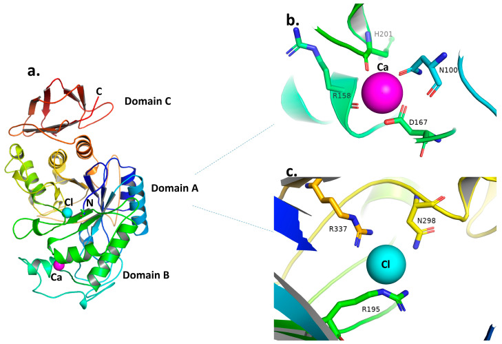
Figure 2.
(a) Ribbon diagram of the structure of human pancreatic α-amylase as a representative for GH13 α-amylase. The three structural domains (A, B, and C) are indicated and colored as follows: Domain A in blues, greens, yellows, and oranges; Domain B in lime green and pale cyan; and Domain C in red. The N and C terminals are colored blue and red, respectively. The calcium and chloride ions are shown as magenta and cyan spheres, respectively. (b) Human pancreatic α-amylase calcium binding site; the sticks represent residues making ligand interactions with calcium, which is represented as a magenta sphere. (c) Human pancreatic α-amylase chloride binding site; the sticks represent residues making ligand interactions with chloride, which is represented as a cyan sphere. From the structure, (a–c) were adopted with PDB entry code 1HNY [33] and produced using PyMol [47].

