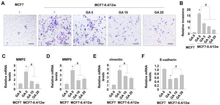
Figure 4.
Low concentrations of gallic acid decreases acidity-induced metastatic characteristics in MCF7-6.4/12w cells. (A,B) Following 48 h of treatment with 5, 10, and 20 μM of gallic acid in MCF7-6.4/12w cells, an invasion assay was conducted (A) and the relative number of invaded cells compared to the normal MCF7 cells (B). (C–F) MCF7-6.4/12w cells were treated with 0, 10, and 20 μM of gallic acid for 48 h, and the mRNA expression levels of MMP2 (C), MMP9 (D), vimentin (E), and E-cadherin (F), and were analyzed using real-time PCR. The relative mRNA expression levels were calculated with the number of untreated normal MCF7 cells as the reference. * p < 0.05 vs. MCF7, # p < 0.05 vs. untreated MCF7-6.4/12w. Scale bar = 100 μm.

