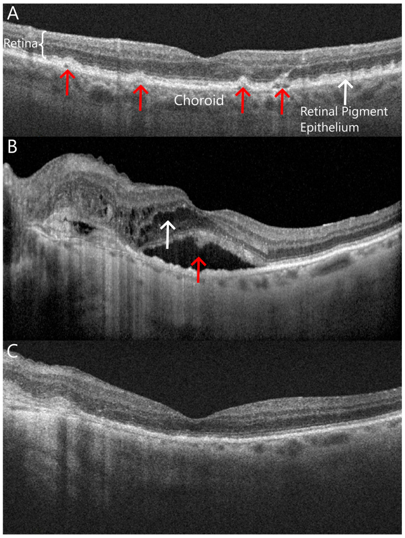
Figure 1.
Optical coherence tomography images show cross-sectional in vivo images of the macula in the eyes in various stages of age-related macular degeneration (AMD). (A) An eye with non-exudative or “dry” AMD with drusen between Bruch’s membrane and the retinal pigment epithelium (red arrows); (B) an eye with active exudative or “wet” AMD with subretinal (red arrow) and intraretinal fluid (white arrow) in the macula; (C) the same eye shown in B after receiving several intravitreal anti-VEGF injections with interval resolution of intraretinal and subretinal fluid.

