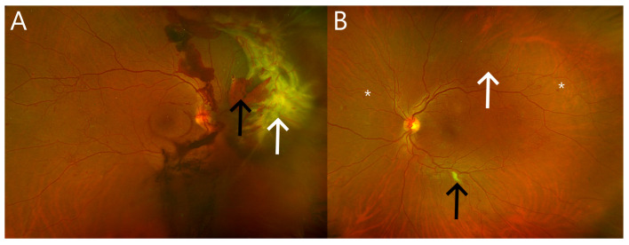
Figure 2.
Wide field pseudocolor retinal images of eyes with proliferative diabetic retinopathy. (A) Right eye with florid neovascularization of the optic disc and retina resulting in tractional retinal detachment (white arrow) and hemorrhage in multiple layers including the vitreous (black arrow). (B) Left eye of the same patient showing less advanced proliferative diabetic retinopathy with less florid retinal neovascularization along the major retinal vessels (white arrow), a cotton wool spot (black arrow), and few dot-blot retinal hemorrhages (asterisks); no vitreous hemorrhage or a tractional membrane has occurred.

