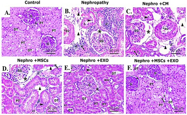
Figure 6.
Effect of Br-MSCs and/or their derived EXOs on the renal histopathological changes (A–F). (A) Photomicrographs of H&E-stained sections showing histological features of control group, showing a spherical renal corpuscle lined by Bowman’s capsule, the glomerulus (G) appears as a large cellular mass surrounded by narrow Bowman’s space (star). Proximal convoluted tubules (PT) have a narrow star-shaped lumen and are lined by acidophilic cuboidal cells with an apical brush border. The distal convoluted tubules (DT) have a wider lumen and are lined by more cells. (B) Photomicrographs of H&E-stained sections showing histological features of the nephropathy group showing marked distorted cortical structures and features of tubule degeneration. The lining epithelium of some tubules (t) is sloughed into their lumina (arrowhead). Other tubules showed vacuolation of the lining epithelium with pyknotic nuclei (bifid arrow). Shrunken glomeruli (SG), wide Bowman’s space (star), and wide spaces between tubules (triangle) are seen. Heavy inflammatory cell infiltration (if) and thick-walled blood vessels (bv) can be noticed between the tubules. (C) Photomicrographs of H&E-stained sections showing histological features of nephropathy+ CM group, showing nearly similar histological findings as in nephropathy group in the form of exfoliated tubular cells (arrowhead), pyknotic nuclei (bifid arrow), shrunken glomeruli (SG), and wide Bowman’s space (star). Also, foci of inflammatory cell infiltration (if), wide spaces (triangle) between the tubules (t), and thick-walled blood vessel (bv) can be noticed. (D) Photomicrographs of H&E-stained sections showing histological features of nephropathy + MSCs group showing some normal cortical tubules having wide lumina (DT) and star-shaped lumina (PT). However, the lining epithelium of affected tubules showed pyknotic nuclei (bifid arrow) and the lumina of some tubules (t) showed exfoliated epithelium (arrowhead). Shrunken glomeruli (SG), wide Bowman’s space (star), wide spaces between tubules (triangle), and inflammatory cell infiltration (if) can be seen in the interstitium. (E) Photomicrographs of H&E stained sections showing histological features of nephropathy + EXOs group showing marked restoration of normal cortical tubules architectures that appeared with wide lumina (DT), star-shaped lumina (PT), and normal glomerular structure (G), surrounded by narrow Bowman’s space (star). However, the lining epithelium of few affected tubules showed dark nuclei (bifid arrow). (F) Photomicrographs of H&E-stained sections showing histological features of nephropathy + MSCs + EXOs group, showing a histological profile comparable to the control group in the form of normal glomerular structure (G), surrounded by narrow Bowman’s space (star). Proximal convoluted tubules (PT) lined by tubular cells have a star-shaped lumen and vesicular nuclei. Distal convoluted tubules (DT) have wider lumen. Only a few distal tubules (t) showed dark nuclei (bifid arrow) (H&EX 400, Scale bar = 50 μm). n = 5–6 rats per group.

